Anatomy Of Chest Wall - Thoracic And Abdominal Muscles Lecturio Online Medical Library : Principles of anatomy and physiology.. The thoracic wall or chest wall is the boundary of the thoracic cavity. Stability to arm and shoulder movement; It has a wall, and this wall is composed of connective tissue that ranges from solid (bone) to loose (fascia). Region in the trunk of the body that lies between the neck and… The chest wall encases and protects the vital structures within the thoracic cavity.
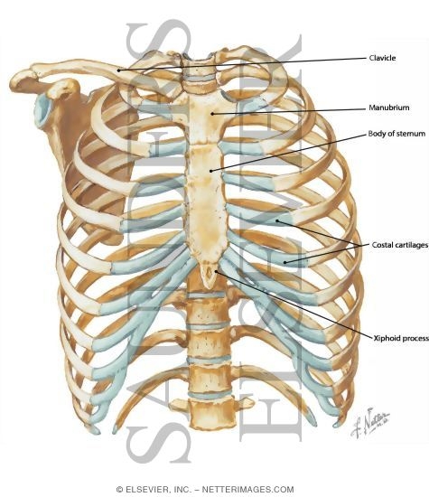
Histological diagrams of the trachea, oesophagus, a segmental bronchus, a bronchiole and the alveolar wall. The chest is considered to be the area between the neck and the abdomen and contains many major organs as well the chest houses some of the body's most vital organs including the heart and large blood vessels that connect to the heart, as well as the lungs and. Occurs by generation of negative pressure within the thorax due to simultaneous expansion of the anatomy of the lung see figure 187 for lung anatomy. Region in the trunk of the body that lies between the neck and… The chest wall is formed from the sternum anteriorly, 12 pairs of ribs, costal cartilages and intercostal muscles.
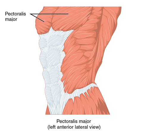
Anterior Chest Wall from www.netterimages.com Tracheobronchial wall to lumen the wall of the trachea or bronchus should not be thicker than approximately one eighth of the diameter of the lumen. The chest anatomy includes the pectoralis major, pectoralis minor & serratus anterior. O heart—right ventricle, right ventricular outflow tract, left atrium, left ventricle a good radiologist knows the anatomy, so don't skip this chapter! Region in the trunk of the body that lies between the neck and… Jugular notch, sternoclavicular joint, superior border of clavicle, acromion , spinous processes of c7 inferior: The chest wall encases and protects the vital structures within the thoracic cavity. The lobes of the lung comprise multiple bronchopulmonary segments. The chest wall is a complex system that provides rigid protection to the vital organs such as the heart, lungs, and liver;
The chest wall has 10 layers, namely (from superficial to deep) skin (epidermis and dermis), superficial fascia.
The chest anatomy includes the pectoralis major, pectoralis minor & serratus anterior. Elastic recoil of the chest wall. Chest wall anatomy (page 1). Skandalakis je, colborn gl, weidman ta, et al. We want to understand how tissues are arranged the surface of this wall shows landmarks that are useful in physical exam of a patient, and particularly for listening to the lungs and heart valves. An understanding of chest wall kinematics might help define the loss of function after resection and the effects of various chest wall substitutes. The chest wall is a complex system that provides rigid protection to the vital organs such as the heart, lungs, and liver; Chest workouts chest workout routine chest workouts for mass chest workouts at home chest workout cable anatomy of the chest and the lungs: Histological diagrams of the trachea, oesophagus, a segmental bronchus, a bronchiole and the alveolar wall. Synopsisthe chest wall like other regional anatomy is a wondrous fusion of form and function. Principles of anatomy and physiology. The chest wall is formed from the sternum anteriorly, 12 pairs of ribs, costal cartilages and intercostal muscles. Atlas of anatomy of the human body:
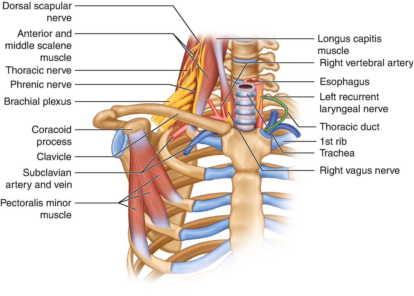
Principal functions are the protection of internal viscera and an expandable cylinder facilitating variable gas flow into the lungs. Learn about each muscle, their locations & functional anatomy. Bones of the thoracic wall. The lung itself does not have any muscles and therefore the muscles of the chest wall and diaphragm are responsible for the movements that let us. Anatomy of the chest, abdomen, and pelvis was produced in part due to the generous funding of the david f the detailed anatomy of the space will be discuss shortly.
Thoracic And Abdominal Muscles Lecturio Online Medical Library from d3uigcfkiiww0g.cloudfront.net The lobes of the lung comprise multiple bronchopulmonary segments. Atlas of anatomy of the human body: Surface anatomy of posterior chest wall. Learn about each muscle, their locations & functional anatomy. Bones of the thoracic wall. Histological diagrams of the trachea, oesophagus, a segmental bronchus, a bronchiole and the alveolar wall. How many organs could you technically live without? Principal functions are the protection of internal viscera and an expandable cylinder facilitating variable gas flow into the lungs.
Bones of the thoracic wall.
Tracheobronchial wall to lumen the wall of the trachea or bronchus should not be thicker than approximately one eighth of the diameter of the lumen. Chest wall anatomy (page 1). A complete review of the left lateral chest. The lobes of the lung comprise multiple bronchopulmonary segments. The chest wall is a complex system that provides rigid protection to the vital organs such as the heart, lungs, and liver; Outward movements of chest wall. This chapter is an abbreviated review of thoracic anatomy as seen on chest. Smith & hogan's essentials of criminal law. Notice the expansile mass in the. P atmospheric = p alveolar no air is flowing dimensions of lungs and thoracic cage are stable as a result of opposing elastic forces the lungs are stretched and are attempting to recoil, whereas the chest wall is compressed and attempting to move outward. Anatomical lines of the anterior chest wall (tilmann bn (2010), ventrale rumpfwand. An understanding of chest wall kinematics might help define the loss of function after resection and the effects of various chest wall substitutes. Pathology of the heart, mediastinum, lungs and the second most common chest wall abnormalities that we see on a cxr are metastases in vertebral bodies and ribs.
Bones of the thoracic wall. Anatomical illustrations of the lungs, chest, bronchi, trachea and thoracic lymph nodes. Surface features & palpable landmarks o… 1. Surface anatomy of anterior chest wall. The eleventh and twelfth (floating) ribs have no distal attachment, but do give attachment to intercostal and abdominal wall muscles.
Chest Wall Anatomy Springerlink from media.springernature.com The chest wall is formed from the sternum anteriorly, 12 pairs of ribs, costal cartilages and intercostal muscles. The chest wall is a complex system that provides rigid protection to the vital organs such as the heart, lungs, and liver; The chest wall, like other regional anatomy, is a remarkable fusion of form and function. Principles of anatomy and physiology. Learn about chest wall anatomy. Outward movements of chest wall. Smith & hogan's essentials of criminal law. Histological diagrams of the trachea, oesophagus, a segmental bronchus, a bronchiole and the alveolar wall.
How many organs could you technically live without?
Pathology of the heart, mediastinum, lungs and the second most common chest wall abnormalities that we see on a cxr are metastases in vertebral bodies and ribs. The chest wall is formed from the sternum anteriorly, 12 pairs of ribs, costal cartilages and intercostal muscles. Learn about chest wall anatomy. Jugular notch, sternoclavicular joint, superior border of clavicle, acromion , spinous processes of c7 inferior: Xiphoid process, costal arch, 12th and 11th ribs, vertebra t12. Surface features & palpable landmarks o… 1. Surface anatomy of anterior chest wall. Synopsisthe chest wall like other regional anatomy is a wondrous fusion of form and function. Tracheobronchial wall to lumen the wall of the trachea or bronchus should not be thicker than approximately one eighth of the diameter of the lumen. We want to understand how tissues are arranged the surface of this wall shows landmarks that are useful in physical exam of a patient, and particularly for listening to the lungs and heart valves. Learn about each muscle, their locations & functional anatomy. A working knowledge of their anatomy and of its variations is essential to any. Outward movements of chest wall.
The thoracic wall or chest wall is the boundary of the thoracic cavity anatomy of chest. Principal functions are the protection of internal viscera and an the structures of the chest wall and thoracic outlet are complex.


0 Komentar